The molecular basis for sarcomere organization in vertebrate skeletal muscle
Wang Z, Grange M, Wagner T, Khoo AL, Gautel M, Raunser S (2021) Cell
Source
Sarcomeres are force-generating and load-bearing devices of muscles. A precise molecular picture of how sarcomeres are built underpins understanding their role in health and disease. Here, we determine the molecular architecture of native vertebrate skeletal sarcomeres by electron cryo-tomography. Our reconstruction reveals molecular details of the three-dimensional organization and interaction of actin and myosin in the A-band, I-band, and Z-disc and demonstrates that α-actinin cross-links antiparallel actin filaments by forming doublets with 6-nm spacing. Structures of myosin, tropomyosin, and actin at ~10 Å further reveal two conformations of the “double-head” myosin, where the flexible orientation of the lever arm and light chains enable myosin not only to interact with the same actin filament, but also to split between two actin filaments. Our results provide unexpected insights into the fundamental organization of vertebrate skeletal muscle and serve as a strong foundation for future investigations of muscle diseases.
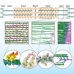
Organisation des Sarkomers auf molekularer Ebene. Erste Zeile zeigt eine schematische Darstellung des Sarkomers. Die zweite Zeile zeigt die dreidimensionale Organisation des Sarkomers und die Plastizität auf molekularer Ebene (dünne Filamente: grün, rosa, a-Actinin: blau). Die dritte Reihe zeigt die Interaktion der Muskelproteine im Detail. Die ersten beiden Blasen zeigen die Interaktion der Myosinköpfe (gelb, orange, rot) mit Aktin (grün). Die dritte Blase v. l. zeigt Details von Aktin, Tropomyosin und Troponin im A-Band. Die letzte Blase (rechts) zeigt das unregelmäßige Geflecht von a-Actinin (blau), das Aktinfilamente (grün, rosa) in der Z-Scheibe vernetzt.
MPI f. molekulare Physiologie
Organisation des Sarkomers auf molekularer Ebene. Erste Zeile zeigt eine schematische Darstellung des Sarkomers. Die zweite Zeile zeigt die dreidimensionale Organisation des Sarkomers und die Plastizität auf molekularer Ebene (dünne Filamente: grün, rosa, a-Actinin: blau). Die dritte Reihe zeigt die Interaktion der Muskelproteine im Detail. Die ersten beiden Blasen zeigen die Interaktion der Myosinköpfe (gelb, orange, rot) mit Aktin (grün). Die dritte Blase v. l. zeigt Details von Aktin, Tropomyosin und Troponin im A-Band. Die letzte Blase (rechts) zeigt das unregelmäßige Geflecht von a-Actinin (blau), das Aktinfilamente (grün, rosa) in der Z-Scheibe vernetzt.
. Die dritte Reihe zeigt die Interaktion der Muskelproteine im Detail. Die ersten beiden Blasen zeigen die Interaktion der Myosinköpfe (gelb, orange, rot) mit Aktin (grün). Die dritte Blase v. l. zeigt Details von Aktin, Tropomyosin und Troponin im A-Band. Die letzte Blase (rechts) zeigt das unregelmäßige Geflecht von a-Actinin (blau), das Aktinfilamente (grün, rosa) in der Z-Scheibe vernetzt.")