Cryo-electron tomography
The Electron Microscopy facility also supports the complete cryo-ET workflow. Two Thermo Scientific Aquilos 2 DualBeam™ cryo-scanning electron microscope allows milling of ultra-thin cryo-lamellas (ca. 80 nm) of cell organelles and isolated cells with a focused ion beam (FIB). Images of these lamellas are acquired at different stage tilts around the tilt axis to produce a 3D volume of the field of view. With subtomogram averaging, the Fisher Scientific Titan Krios instruments are able to resolve high resolution protein structure maps below 3 Å resolution.
Furthermore, a plasma focused ion beam (PFIB) DualBeam™ Thermo Scientific Hydra CX instrument permits milling of much larger volumes than the standard FIB. This enables the investigation of not only isolated cells but also multicellular tissues or organoids. For the vitrification of these thick samples, the high pressure freezer (Leica EM ICE or Leica HPM100) is used. Due to thickness limitations during high pressure freezing (< 200 μm) it is necessary to cut sections with a microtome provided by the facility, e.g. Leica vibratome VT1200S, Leica cryostat CM3050S or Leica cryo ultra-microtome EM FC6.
FIB milling porcess of a muscle fibre
A general challenge in cryo-ET is the targeting of specific biological structures (regions of interest) during the FIB milling process. A stand-alone cryo-fluorescence light microscope (FEI CorrSight) and fluorescence light microscopes integrated into both Fisher Scientific Aquilos instruments (Delmic METEOR system) help to correlate with a fluorescence signal during the milling process. These regions of interest in the vitrified biological sample are therefore labeled beforehand with a specific fluorescence marker.
Zooming in on Muscle Cells
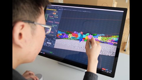
Watch how an international Team led by Prof. Stefan Raunser has produced the first high-resolution 3D image of the sarcomere, the basic contractile unit of skeletal and heart muscle cells, by using electron cryo-tomography (cryo-ET).
