Structure of key protein for cell division puzzles researchers
Max Planck Institute’s researchers provide a first 3D snapshot of the CCAN protein complex and raise fundamental questions towards the creation of artificial chromosomes
Human cell division involves hundreds of proteins at its core. Knowing the 3D structure of these proteins is pivotal to understand how our genetic material is duplicated and passed through generations. The groups of Andrea Musacchio and Stefan Raunser at the Max Planck Institute of Molecular Physiology in Dortmund are now able to reveal the first detailed structure of a key protein complex for human cell division known as CCAN. By using cryo-electron microscopy, the researchers show important features of the complex’s 16 components and challenge previous assumptions about how the complex is able to recognize the centromere, a crucial region of chromosomes in cell division.
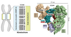
. Right: Model of the surface of the CCAN’s 16 components organized in different subcomplexes.")
At the centre of cell division
The centromere is a constriction in the chromosome, made of DNA and proteins. Most importantly, the centromere is the dock for the kinetochore, a machinery of about 100 proteins that drives the separation of two identical chromosomes during cell division and their delivery to the daughter cells. Previous research has shown that the kinetochore docks onto the centromere through the CCAN complex: The CCAN interacts with the centromere protein A, the landmark protein of the centromere. CCAN is also responsible for replenishing the centromere protein A once the cell division has taken place. Yet, the details of the interaction between CCAN and the centromere protein A remain elusive. “Understanding how CCAN recognises and binds to the centromere could potentially lead us to build a centromere from scratch”, says Musacchio. The centromere is a major hurdle for synthetic biologists who aim to engineer artificial chromosomes to restore missing functions or introduce new ones in cells.
Unsolved questions at the core
Scientists identified the CCAN complex over 15 years ago. “Yet, building up a pipeline to synthesize all proteins in vitro has been a major obstacle”, says Musacchio. After obtaining a first reconstitution of the human CCAN complex in vitro, Musacchio joined forces with Stefan Raunser, also at MPI Dortmund, who applied cryo-electron microscopy on the whole CCAN protein complex.
In the new publication, the MPI groups have been able to determine the 3D structural details of the human CCAN complex, highlighting its unique features and the implications for an interaction with the centromere protein A. “Contrary to what was expected, this structure does not directly recognise the centromere protein A in the standard configuration”, says Musacchio. The centromere protein A is most commonly packed with DNA and other proteins as a nucleosome, the standard unit of the genetic material. The authors are now suggesting that the centromere protein A may be embedded in the centromere with a different configuration that may facilitate the crucial interaction with CCAN. They plan to identify conditions that could lead to this new configuration and prove their hypothesis.